
What a 3D CBCT Scan Reveals That Standard X-Rays Miss Entirely
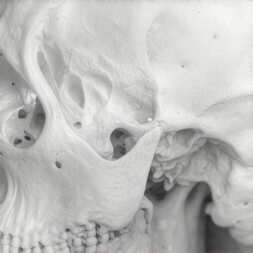
What a 3D CBCT Scan Reveals That Standard X-Rays Miss Entirely
As a Registered Dental Hygienist and a Medit Champion, I’ve spent years looking at the intricate landscapes of the human mouth. For decades, the gold standard for diagnostic imaging was the traditional 2D X-ray. While these “flat” films are incredibly useful for spotting cavities between teeth, they have a significant limitation: they attempt to represent a three-dimensional human being in only two dimensions. It is like looking at a flat paper map of the Blue Ridge Mountains; you can see the roads, but you have no real sense of the elevation, the hidden ravines, or the depth of the forest. In modern dentistry, specifically when visiting a dentist in culpeper va, we now have access to 3D drone footage of your anatomy. This technology is known as CBCT (Cone-Beam Computed Tomography). By capturing a 3D volume of data, we can see through bone and tissue to identify issues that traditional X-rays miss entirely, ensuring that your primary care dentistry is as precise as possible.
The Technical Leap: 2D vs. 3D Imaging with a Dentist in Culpeper VA
To understand why all smiles dentistry has transitioned toward 3D imaging, we have to look at the mechanics of the technology. A standard dental X-ray – whether it’s a bitewing or a periapical – is a static snapshot. The radiation passes through your cheek and teeth to hit a sensor on the other side. Because the image is flat, structures often overlap. A tooth root might be hiding a small infection behind it, or a dense area of jawbone might obscure a tiny fracture. This “shadowing” effect is the primary cause of undiagnosed dental pain.
When you visit a culpeper dentist for a 3D scan, the experience is fundamentally different. The CBCT machine features a C-arm or gantry that rotates 360 degrees around your head. During this single rotation, it captures hundreds of high-resolution images from every conceivable angle. These “slices” are then reconstructed by advanced software into a single 3D model. As a Medit Champion, I find this technology revolutionary because it allows us to virtually “walk through” your jawbone. We can rotate the image, peel away layers of bone, and view your teeth from the inside out. This level of detail is essential for “Family Dentistry Secrets: Maintaining Oral Health with Modern Techniques,” as it moves us from reactive treatment to proactive prevention.
For patients seeking dentists culpeper va, this means higher diagnostic accuracy. Whether we are evaluating a child’s developing palate or planning a complex adult restoration, the 3D view eliminates the guesswork that used to be inherent in 2D films. It provides a comprehensive map of the dental landscape, including soft tissues, nerve paths, and bone density, all in one scan.
5 Critical Things Standard X-Rays Miss: Why All Smiles Dentistry Uses 3D
In my clinical experience, I have seen numerous cases where a patient complained of persistent discomfort, yet their 2D X-rays looked “perfect.” This is where the 3D CBCT scan becomes a lifesaver. Here are five specific things that standard X-rays frequently miss.
1. Hidden Root Canals
Human anatomy is rarely textbook-perfect. Many teeth, particularly the maxillary first molars, often have a “hidden” fourth canal known as the MB2. On a 2D X-ray, this canal is almost always obscured by the larger canals surrounding it. If a dentist performs a root canal but misses this tiny, hidden passage, bacteria will remain trapped inside. This leads to a “failed” root canal and chronic pain. By using 3D imaging at allsmilesdentalculpeper.com, we can see the exact number and shape of every canal before the procedure even begins. If you’ve ever wondered about “The ‘Sip Test’ to Determine if Your Tooth Sensitivity is Nerve-Related,” a 3D scan is often the final piece of that diagnostic puzzle.
2. Fine Root Fractures
Micro-cracks or vertical root fractures are the “ninjas” of dental problems – they are incredibly hard to see and cause immense pain. A 2D X-ray only shows a fracture if the X-ray beam happens to pass exactly parallel to the crack. Since most cracks are microscopic and angled, they stay hidden. A 3D scan allows us to view the tooth in cross-sections of less than a millimeter. This precision helps us identify “Why Your Crown Feels Like It Has a High Spot Only When You Grind,” which is often actually a sign of a deep-seated fracture that a 2D film would never reveal.
3. Nerve Pathway Mapping
Safety is paramount in primary care dentistry. When extracting wisdom teeth or placing implants, we must know exactly where the inferior alveolar nerve is located. In a 2D image, the nerve might look like it’s a safe distance from the tooth root, but in reality, the root could be wrapped right around it. A 3D scan provides the “z-axis” – the depth – showing us the exact spatial relationship between your teeth and your nerves. This prevents permanent numbness and surgical complications.
4. Bone Volume and Density
If you are considering dental implants, a 2D X-ray is insufficient. It can tell us the height of the bone, but it cannot tell us the width or the quality. You might have plenty of height but bone that is “paper-thin,” which would lead to implant failure. This technology is vital for identifying “The Sign that you need a bone graft before getting a dental implant.” By assessing bone density in 3D, we ensure the foundation is strong enough to support your new smile.
5. Sinus Proximity
The roots of your upper teeth often sit very close to your maxillary sinuses. In some cases, the roots actually extend into the sinus cavity. A 2D X-ray makes it look like the teeth and sinuses are on the same plane, but it doesn’t show the thickness of the sinus floor. 3D imaging allows your oral care dentist to see the exact proximity, which is crucial for preventing sinus perforations during extractions or 24hr dental care emergencies.
Safety First: Debunking the Radiation Myth
One of the most common questions I hear as a hygienist is: “Is a 3D scan safe?” It is a valid concern, but it is important to put the numbers into perspective. While a CBCT scan does involve more radiation than a single 2D bitewing, it is significantly safer than a traditional medical CT scan. In fact, a dental 3D scan typically delivers a radiation dose that is roughly equivalent to a few days of natural background radiation from the environment.
At all smiles dentist, we follow the ALARA principle: As Low As Reasonably Achievable. We don’t take 3D scans for every prophy dentistry visit. Instead, we use them as a “sharper, more accurate diagnostic” tool when the clinical situation warrants it. By getting a clear, 3D picture at the start, we often avoid the need for multiple follow-up 2D X-rays and “exploratory” surgeries. This actually reduces the total radiation exposure and the overall cost of care over the long term. When you choose a oral care dentist who uses this tech, you are choosing precision over guesswork.
Why All Smiles Dentist Invests in Advanced CBCT Technology
You might wonder why every dentist culpeper va doesn’t have this technology. The truth is, CBCT machines are a massive investment in both equipment and training. However, culpepper family dentistry believes that providing the best dentistry requires the best tools. We aren’t just looking for cavities; we are looking at the health of your entire craniofacial structure.
Our investment in 3D imaging reflects our commitment to “millimeter precision.” This is especially true for our dentistry services involving dental implants and orthodontics. Research by experts like Dr. Sanie has shown that 3D guided implant surgery has a significantly higher success rate and faster healing time than traditional methods. By knowing exactly where the bone is densest and where the nerves are located, we can place implants with a level of accuracy that was impossible ten years ago. We also utilize this tech to explore “The Hidden Benefits of Invisalign for Family Dental Care in 2025,” as 3D imaging helps us predict how teeth will move through the bone more effectively than 2D ever could.
The Patient Experience: What to Expect During Your Scan
If your prophy dentistry provider recommends a 3D scan, there is no need for anxiety. The process is entirely non-invasive and much faster than most people expect. Unlike a medical CT where you have to lie down in a “tube,” a dental CBCT scan is open. You will usually stand or sit in the center of the machine.
A staff member will help with “proper patient positioning” to ensure the image is crystal clear. You’ll be asked to stay very still for about 10 to 20 seconds while the machine’s arm makes one quiet revolution around your head. There is no “closed-in” feeling, and nothing goes inside your mouth during the scan itself. It is a quick, painless part of our comprehensive allsmilesdentalculpeper.com experience. Whether we are preparing for allsmilesdentalculpeper.com or a routine check-up, this scan provides the peace of mind that nothing is being overlooked.
Conclusion: Is a 3D Scan Right for You?
While a 3D scan isn’t necessary for every single prophy dentistry appointment, it has become the gold standard for modern dental care. If you are experiencing unexplained pain, planning for dental implants, or need a complex extraction, the 3D CBCT scan is the only way to ensure your dentist has the full picture. It reveals the hidden canals, the micro-fractures, and the complex nerve pathways that standard X-rays miss entirely.
At all smiles dentistry, we are proud to offer this technology to the Culpeper community. We believe that better imaging leads to better diagnoses, more conservative treatments, and ultimately, healthier smiles. If you are looking for a oral care dentist who prioritizes technology and patient safety, we invite you to visit allsmilesdentalculpeper.com to schedule your consultation. Don’t leave your oral health to chance – see the difference that 3D precision can make.